Brain (cont'd)
The Lateral Geniculate Nuclei
Neuroscientists commonly describe the thalamus (a gray matter nucleus deep within the brain) as a relay station between in-coming sensory information and the cortex. And yet, whenever they go on to describe, say, axonal projections from the eye, they almost never mention the thalamus as the thalamus. They refer instead to the lateral geniculate nucleus, which is, in fact, that particular portion of the thalamus to which ganglion cell axons of the retina project. The cells of the lateral geniculate nucleus project, in turn, as promised, to the primary visual cortex (also known as V1 or the striate cortex).
P and M Pathways
P cells, some 80% of the population of the retinal ganglion cells, terminate in the parvocellular layers of the lateral geniculate nucleus. M cells, a mere 10%, terminate in the magnocellular layers.
P cells can be connected to cones in such a way that they are excited by some wavelengths of light and inhibited by others--for which reason, they are often described as color-opponent.
M cells, on the other hand, respond indescriminently to wavelengths of varying illumination and, as a consequence, are referred to as broadband.
M and P cells travel largely discrete, parallel pathways from the retina to their various nuclear destinations within the brain. For once, to simplify matters, anatomists have agreed to give these natural, obvious monikers: the M pathway and the P pathway.
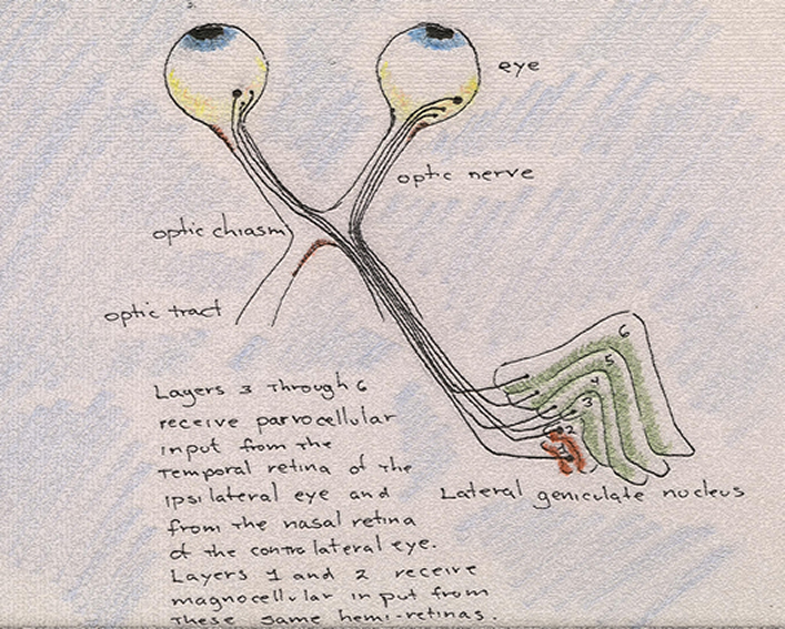
When stained, the six laminae of the lateral geniculate nucleus, are easy to discern: 4 are parvocellular, 2 magnocellular. The latter are the ventral-most layers.
The organization or arrangement of the layers of the lateral geniculate nucleus and of the axons that they receive is complicated by the fact that the contralateral eye (the right eye, for example, if we are speaking of the left lateral geniculate nucleus) projects to layers 1, 4 and 6, while the ipsilateral eye (the left eye if we are again speaking of the left lateral geniculate nucleus) projects to layers 2, 3 and 5.
An important and curious fact regarding the axon projections of retinal ganglion cells is that they maintain their positions relative to one another as they pass from the retina to the lateral geniculate nucleus to the primary visual cortex. In other words, they project a map of the retina onto all subsequent intercranial destinations within the brain, a map thus designated as retinotopic. However, this map is free of the crabbed, concave confines of the retina, and density, therefore, can be translated as space. The result is a map in which the fovea occupies a larger amount of space than it does in the retina. This is because the fovea is the densest portion of the retina; when spread out, it reveals how much more information is concentrated in the center of the eye than in its periphery.
Primary Visual Cortex (aka, V1 and the Striate Cortex)
[Our apologies as this section is still to be edited]